

Multimodal blood perfusion phantom system
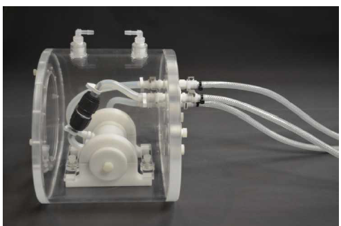
Multimodal blood perfusion phantom system |
|
| description: | Programmable multi-modal dynamic contrast enhancement (DCE) blood perfusion phantom through Based on step function or typical clinical arterial input function (AIF) input Pressure controls blood flow changes to simulate extracorporeal blood flow and two |
|---|---|
Programmable multi-modal dynamic contrast enhancement (DCE) blood flow perfusion phantom simulates extracorporeal blood flow and two-chamber contrast blood flow by controlling blood flow changes based on step function or typical clinical arterial input function (AIF) input pressure Kinetics This innovative phantom can generate a wide range of predictable, reproducible and quantifiable time-concentration curves (TCC). These curves generate a wide range of real input and output functions that simulate clinically relevant perfusion TCC. The phantom generation has The ability of the DCE quality assurance protocol for real blood flow provides an excellent model for the verification of perfusion and kinetic modeling, and enables DCE imaging to be used as a quantitative imaging tool for scanners in contrast imaging mode, or Compatible with CT MRI and PET modes, and contrast imaging mode. The standard DCE phantom prediction software accurately and repeatably predicts the TCC that will be presented when the phantom is scanned to obtain the established input parameter selection and can generate a wide range of physiologically relevant enhancements. Curve, the user can define the required TCC and calculate the input parameters required for the model. The easy-to-use DCE phantom prediction software allows users to select the following: CT MR or PET imaging method Maximum time injection parameter output flow and perfusion pump flow.
Applications
CT
-Perfusion/DCE CT
-Image noise and partial volume effect quantification
-CT digital linearity and sensitivity
MR
-Based on the size and phase of DCE-MR
-Arterial Spin Labeling (ASL)
Dynamic PET
Compare various single-mode scanners
Comparison of multi-modal scanners
Kinetic model verification
Assess the interaction between dynamic contrast blood flow and dynamic image acquisition
Features
Product advantages
CT
-Perfusion/DCE CT
-Image noise and partial volume effect quantification
-CT digital linearity and sensitivity
MR
-Based on the size and phase of DCE-MR
-Arterial Spin Labeling (ASL)
Dynamic PET
Compare various single-mode scanners
Comparison of multi-modal scanners
Kinetic model verification
Assess the interaction between dynamic contrast blood flow and dynamic image acquisition
Multimodal DCE blood flow phantom
Compatible with X-ray computed tomography (CT), magnetic resonance (MR) and positron emission tomography (PET) imaging modes
Create a wide range of physiologically relevant time-concentration curves under physiological and steady-state flow rates (TCCs), similar to those observed in clinical perfusion CT/MR
Accurate and highly repeatable comparative measurement in dynamic settings
Predictable and reproducible time enhancement curve
Perform CT digital linearity and sensitivity test, convert CT digital enhancement into concentration DCE prediction software
The easy-to-use automated DCE phantom prediction software for CT MRI and PET imaging produces a wide range of clinically relevant and reproducible contrast enhancement curves
Model data is easily exported to MATLAB data file or Excel spreadsheet
Provide baseline comparison calibration curve.
Provide comparison of real imaging modes of CT, MRI and PET scanners,
Provides the gold standard across multiple imaging platforms
Develop dynamic collection protocol to improve dose efficiency
Improve quantitative contrast enhanced imaging and quantify scanner variability
Use the time decay curve generated by phantom
Evaluate the sensitivity of a single scanner
For different CT scanners and acquisition protocols
Perform a basic ROI sensitivity analysis test
Further verify the parameters of the kinetic model
Torso Shell, made of acrylic and sealed
Fluid Mass Exchange Cylinder which embodies a spiraling interior perforated distribution tube with input and output tubes
Injection Port complete with backflow prevention, syringe port and required fittings for typical clinical IV administration.
Needle Valves Control (2)
-4” (6.35 mm) ID carbon stecl for CT/PET
-44" (6.35 mm) ID polypropylene for MRIT
urbine Flow Meters (2):
-50-500 ml/min range (1/8" ID or 3.175 mm)
Turbine Flow Meter Power Supplies (2)
3-Way Ball Diverter Valve:
14" ID, brassTubing-14 feet (4.2672 m), %" (6.35 mm) ID high pressure clear PVO tubing, food grade
-3.70 feet (113 cm), 1/8" (3.175 mm) ID clear PVC tubing, chemical resistant
-5 feet, (152.40 cm), 4" (6.35 mm) high temperature silicone soft rubber tubing.

 Scan QR Code & Subscribe