

logo
×

PRODUCTS

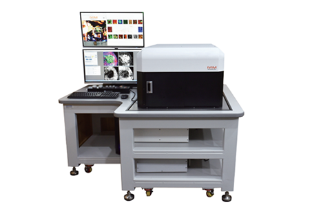
IVM-CM small animal in-situ dynamic analysis imaging system
  • You Are Here :HOME > PRODUCTS > IVM-CM small animal in-situ dynamic analysis imaging system >
  • IVM-CM small animal in-situ dynamic analysis imaging system

    description: In vivo imaging technology mainly uses a set of very sensitive optical detection equipment that can directly monitor cell activities and gene behaviors in living organisms. Through this system, biological processes such as the growth and metastasis of tum
    Technical Features of IVM-CM:
    Two-photon laser wavelength range: 690 nm–1050 nm
    Field of View (FOV): 100×100 µm² to 10×10 mm²
    Imaging resolution: 100 fps @ 512×512 pixels
    Imaging depth: up to 1000 µm
    In vivo imaging targets: liver, lymph nodes, spleen, skin, retina, lung, brain, colon, pancreas, small intestine, prostate, kidney, heart, trachea, esophagus, bone marrow, thymus, etc.

    The all-in-one intravital microscopy system from IVIM Technology is equipped with an ultra-fast rotating polygon mirror scanner, enabling ultra-high-speed intravital imaging (up to 100 fps @ 512×512 pixels). It ensures uniform excitation illumination across the entire field of view (FOV), with no fluorescence signal or signal-to-noise ratio (SNR) reduction in the center, and no excessive photobleaching at the edges. The system delivers consistently high SNR throughout the FOV without the need for excessive photon input to improve image quality.

    Application Fields:
    Immunology research
    Cancer research
    Targeted therapy research
    Molecular pathology
    Application Examples

    Objective
    To apply cranial window-based intravital brain imaging technology for brain tumor studies.
    Result
    Glioma cells were implanted into the brain, and tumor progression was monitored in the same imaging region of the same mouse model for over 15 days. Using IVIM’s intravital microscopy, an increase in the number of glioma cells with rounded morphology was observed as the tumor grew over time.

    Objective
    To perform longitudinal in vivo monitoring of abdominal organs such as the pancreas, spleen, and kidneys using an abdominal imaging window.
    Result
    Using MIP-GFP technology, the dynamic behavior of endogenous monocytes and dendritic cells in the spleen was visualized in real time, along with the dynamic changes of pancreatic islet β-cells. The abdominal imaging window enabled long-term monitoring of cellular dynamics and development.

    Objective
    To monitor and analyze tumor drug efficacy in vivo, including quantitative analysis of angiogenesis, vasodilation, and real-time blood flow changes over time in a single mouse model.
    Result
    Tumor cells were implanted using IVIM technology, and tumor progression was monitored in real time. Long-term studies of drug delivery and distribution targeting breast cancer were conducted using the system.
    Detailed Specifications