

logo
×

PRODUCTS

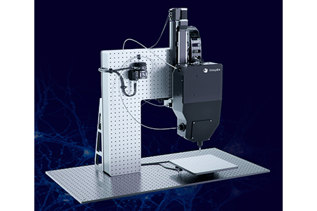
Miniaturized fluorescence imaging system for in vivo animals
  • You Are Here :HOME > PRODUCTS > Miniaturized fluorescence imaging system for in vivo animals >
  • Miniaturized fluorescence imaging system for in vivo animals

    description: This system is an in vivo deep brain imaging platform based on a single ultra-thin multimode optical fiber probe, designed for high-resolution observation of neuronal structures and activity in living animals. It integrates laser-scanning fluorescence mi
    Basic Information
    This system is an in vivo deep brain imaging platform based on a single ultra-thin multimode optical fiber probe, designed for high-resolution observation of neuronal structures and activity in living animals.
    It integrates laser-scanning fluorescence microscopy with digital holographic wavefront shaping, enabling minimally invasive imaging of deep brain regions through a fiber probe on the order of ~100 μm in diameter.
    The platform provides submicron spatial resolution within a small field of view and supports real-time functional imaging of fast neural signals such as calcium and voltage dynamics. It also enables random-access scanning and dynamic focus control for flexible interrogation of neural circuits.
    The system is primarily used in neuroscience research, including studies of deep brain circuit function, disease mechanisms, and pharmacological screening.
    Product Advantages:
    Minimally invasive imaging: Uses an ultra-thin (~100 μm) optical fiber probe, significantly reducing tissue damage.
    Access to deep brain regions: Enables imaging of deep neural structures beyond the reach of conventional optical microscopy.
    High spatial resolution: Achieves submicron resolution, enabling visualization at single-cell and subcellular levels.
    Real-time neural signal recording: Supports fast functional imaging of neural activity, including calcium and voltage signals.
    Flexible scanning capability: Enables random-access scanning for efficient and targeted data acquisition.
    High compatibility with labeling methods: Compatible with various fluorescent indicators and neural probes.
    Suitable for chronic in vivo studies: Designed for long-term imaging and tracking of neural circuit dynamics.
    Workflow

    Main Product Features:
    ·  Single-fiber probe architecture: Uses an ultra-thin multimode optical fiber as the sole optical pathway for deep brain access.
    ·  Digital holographic wavefront control: Enables precise light field reconstruction and focusing at the fiber output.
    ·  Laser-scanning fluorescence imaging: Provides high-contrast fluorescence signal acquisition via point-scanning mechanisms.
    ·  Minimally invasive deep brain access: Allows imaging of deep neural structures with reduced tissue damage.
    ·  High-resolution imaging performance: Achieves submicron spatial resolution for fine structural mapping.
    ·  Real-time functional imaging: Supports fast neural activity measurements such as calcium and voltage imaging.
    ·  Random-access scanning mode: Enables rapid, targeted imaging of selected regions for higher efficiency.
    ·  Miniaturized probe design: Suitable for in vivo small-animal studies and chronic implantation experiments.

    Application Areas:
    1. Structural imaging
    Enables high-resolution visualization of deep brain tissue architecture, including neuronal morphology, axonal projections, and local microstructural organization, based on single-fiber imaging capability.
    2. Calcium imaging
    Uses calcium-sensitive fluorescent indicators to record real-time neuronal population activity, enabling analysis of firing patterns, network synchrony, and functional connectivity dynamics.
    3. Voltage imaging
    Employs voltage-sensitive dyes or indicators to capture millisecond-scale neuronal electrical activity, allowing observation of fast spiking events and transient membrane potential changes.
    4. Blood flow tracking
    Tracks microvascular hemodynamic changes using fluorescence or scattering signals, enabling study of neurovascular coupling between neuronal activity and cerebral blood flow.
    Detailed Specifications