
logo
×

PRODUCTS

Deformability cytometry
  • You Are Here :HOME > PRODUCTS > Deformability cytometry >
  • Deformability cytometry

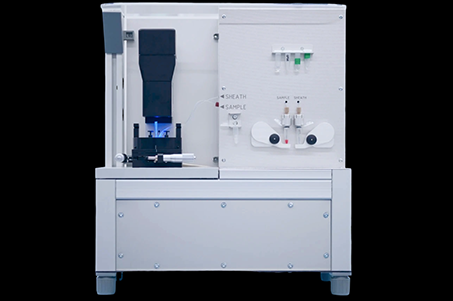
    description: Naiad is a standalone device for researchers who need a tool to conduct deformability cytometry for the physical phenotyping of cells. Naiad is designed for high-speed imaging of cells as they flow through a microfluidic channel. It guarantees a frame ra
    Product Summary
    Naiad is a standalone device for researchers who need a tool to conduct deformability cytometry for the physical phenotyping of cells.
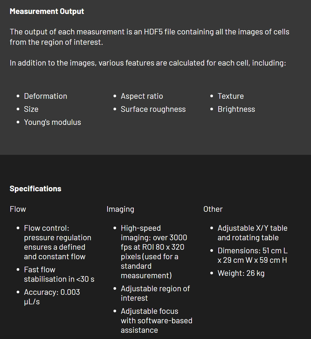
    Naiad is designed for high-speed imaging of cells as they flow through a microfluidic channel. It guarantees a frame rate higher than 3000 frames per second and maintains a stable and quantifiable flow rate, allowing for accurate and reliable cell analysis. Naiad is ideal for day-to-day research projects or long-term clinical studies.
    CytoShot software is used to operate the Naiad from a Windows or Mac computer.
    Advantage:
    We produce devices and software for the analysis of physical properties of cells, including their stiffness, size, shape and morphological features.
    Why do the mechanical properties of cells matter?
    Cell mechanics and other physical properties of cells reflect physiological and pathological changes in cell function. They are promising biomarkers for disease diagnosis and prognosis. For example, we recently found dramatic changes of blood cell mechanics during COVID-19.
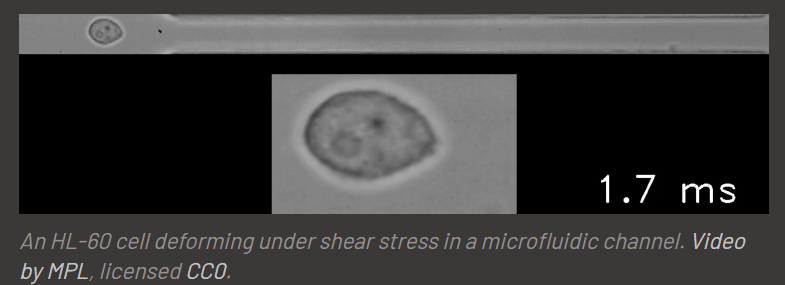
    An HL-60 cell deforming under shear stress in a microfluidic channel. Video by MPL, licensed CC0.
    What is physical phenotyping of cells?
    Physical phenotyping involves the characterization of single living cells by their physical and morphological features such as size, shape and stiffness. An efficient technique for doing this is deformability cytometry. As a label-free method based purely on brightfield images of cells, deformability cytometry adds a new functional and unbiased dimension to flow cytometry. Combined with machine learning, deformability cytometry is a discovery machine for characterizing cell populations and functional states that are invisible to marker-based techniques.

    What is deformability cytometry?
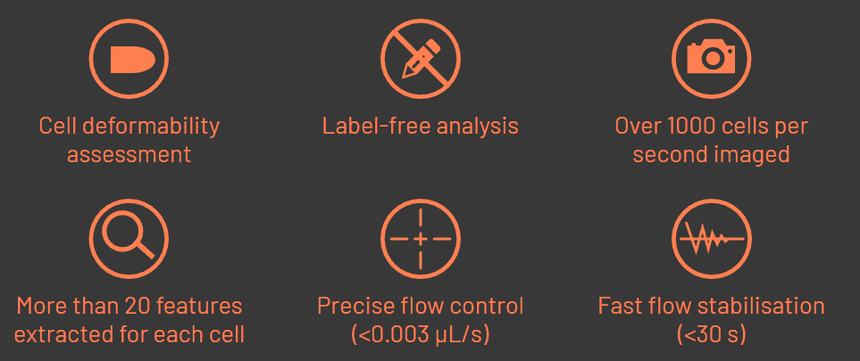
    In deformability cytometry, thousands of single cells flow quickly through a narrow microfluidic channel, where the cells deform due to the forces applied. An image of each cell is taken and analyzed. Cell deformability and other parameters describing the cell's physical phenotype are extracted from the image. Thanks to high-speed video microscopy, our analysis rates of 1000 cells per second approach those of conventional fluorescence-based flow cytometers. Hundreds of thousands of cells are measured within minutes.
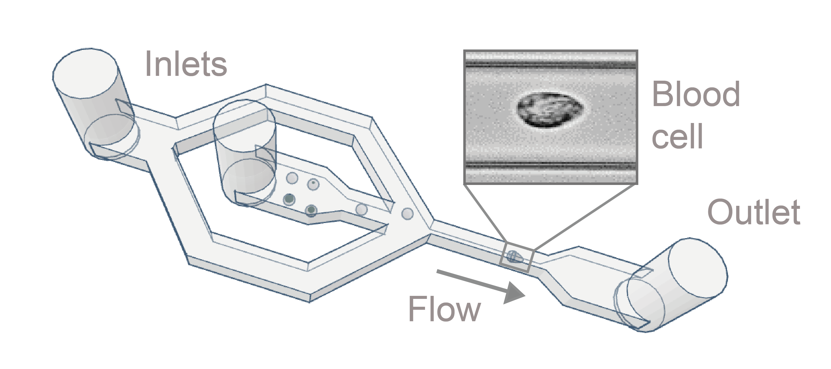
    Scheme of the microfluidic chip used for deformability cytometry
    Which samples can be analyzed?
    Any cell suspension can be measured with deformability cytometry - blood and other liquid biopsies, cells from solid tissues, and cultured cells. All of this is possible without time- and material-consuming sample preparation such as staining. As such, deformability cytometry supports diverse applications in biology, biotechnology and medicine.

    Main character:

    Application

    Technical parameter

    Detailed Specifications