Discoverer™ Brillouin Microscope
Discoverer™ Brillouin Microscope
|
| description: |
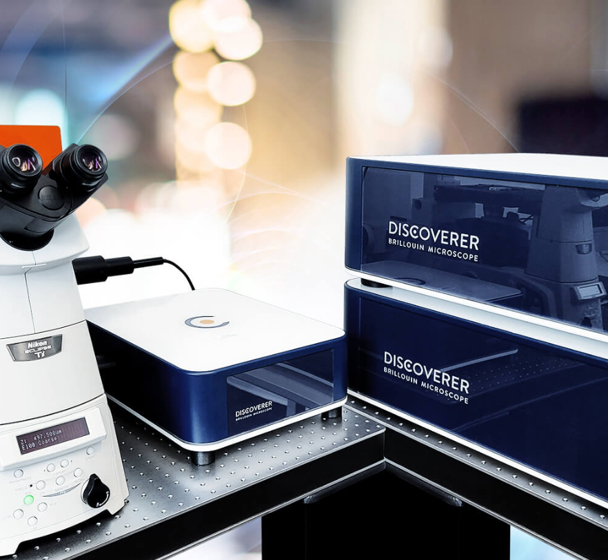
Discoverer系统是一款全新开发的电动倒置或正置研究显微镜附加组件。它兼容蔡司、徕卡、奥林巴斯/Evident 和尼康等主流显微镜品牌。安装快捷简单,只需几分钟即可完成。OptiBridge™ 模块可连接到显微镜机身的侧端口,与系统无缝集成。Discoverer软件充分利用和控制电动显微镜机身,并通过机械成像通道增强光学显微镜的功能,实现真正的多模态成像。Discoverer由经验丰富的机械生物学仪器专业人士开发,可立即使用。 |
Basic Information
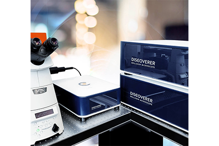
The Discoverer™ system is a newly developed add-on for motorized inverted or upright research microscopes. It is compatible with major microscope brands, including Zeiss, Leica, Olympus/Evident, and Nikon. The installation is quick and straightforward, taking only minutes to complete. The OptiBridge™ module from CellSense connects to a side port of the microscope body, seamlessly integrating the system. The motorized microscope body is fully utilized and controlled via the Discoverer™ software, enhancing the light microscope’s capabilities with a mechanical imaging channel for true multimodal imaging. Developed by experienced professionals in mechanobiology instrumentation, the Discoverer™ is ready to use..
The Discoverer™ is designed for maximum mechanical and thermal stability, compatible with both inverted and upright microscopes for long-term experiments on living samples. It features the latest Brillouin technology, including a multi-stage interferometric spectrum detector, optimized beam path, high-quality optical components with perfected coatings, and innovative automation features. The Discoverer™ is a masterpiece of engineering. Its revolutionary software design enables users to obtain quick results without being experts. It offers a wide range of accessories, modes, and features, providing unparalleled flexibility for any application.
The Discoverer™ system is a newly developed add-on for motorized inverted or upright research microscopes. It is compatible with major microscope brands, including Zeiss, Leica, Olympus/Evident, and Nikon. The installation is quick and straightforward, taking only minutes to complete. The OptiBridge™ module from CellSense connects to a side port of the microscope body, seamlessly integrating the system. The motorized microscope body is fully utilized and controlled via the Discoverer™ software, enhancing the light microscope’s capabilities with a mechanical imaging channel for true multimodal imaging. Developed by experienced professionals in mechanobiology instrumentation, the Discoverer™ is ready to use.
 Product Advantages:
High-resolution and label-free imaging
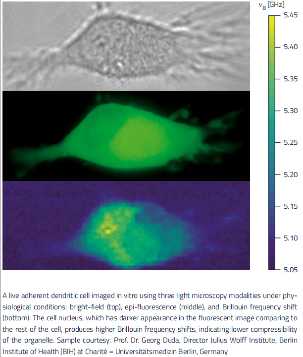
Utilizing Brillouin microscopy technology, it enables non-invasive, label-free 3D mechanical property measurements of living cells, organoids, tissues, and small organisms.
Automation and ease of use
Employs intelligent workflow software such as FlowPlanner™ and PerfectOverlay™ to simplify operation, allowing users with little to no Brillouin microscopy experience to quickly obtain high-quality data.
Multimodal imaging integration
Seamlessly integrates with major inverted or upright microscopes, enabling simultaneous use with brightfield, phase contrast, DIC, fluorescence, and confocal imaging to perfectly combine mechanical information with optical imaging.
Strong compatibility with environmental control
Supports various environmental control devices, suitable for long-term live cell imaging experiments to ensure samples are measured under optimal conditions.
High sensitivity and stability
Uses a low-phototoxicity 780 nm near-infrared laser to minimize sample damage; the system is designed with mechanical and thermal stability for long-term experiments.
Powerful automation features
Includes automated calibration, autofocus, and large-area mapping (MultiMap™), reducing human error and improving data reliability and experimental efficiency.
Compatible with major microscope brands
Fits with mainstream microscopes such as Zeiss, Leica, Nikon, and Olympus, making it easy to integrate into existing experimental platforms.
Main Product Features:
1. Label-Free, Non-Invasive Imaging
Utilizes Brillouin scattering to measure mechanical properties without fluorescence labeling or damaging live samples.
2. 3D Mechanical Imaging Capability
Offers high-resolution 3D mechanical mapping of cells, organoids, tissues, and small organisms.
3. Highly Automated and User-Friendly
Equipped with intuitive software like FlowPlanner™ and PerfectOverlay™, enabling easy setup and operation—even for beginners.
4. Strong Multimodal Compatibility
Seamlessly integrates with brightfield, fluorescence, DIC, and confocal imaging for correlated optical and mechanical data.
5. Environmental Control Support
Compatible with temperature, CO₂, and humidity control systems for long-term live imaging applications.
6. High Sensitivity and Low Phototoxicity
Uses a 780 nm near-infrared laser to minimize photodamage, ideal for long-term observation of sensitive samples.
7. Advanced Data Processing and Visualization
Includes powerful features such as data fitting, edge detection, 3D rendering, and cross-sectional analysis, with batch processing support.
8. Modular Design with Broad Microscope Compatibility
Easily integrates with leading inverted and upright microscopes from Zeiss, Nikon, Leica, and Olympus, allowing easy system upgrades.
Application Areas:
1. 3D Cell Culture & Mechanobiology
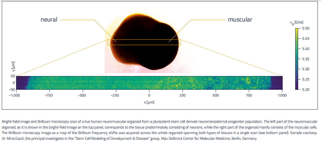
Non-invasive, label-free measurement of the mechanical properties of spheroids and organoids.
Real-time monitoring of tumor organoids (e.g., MCF‑7 breast cancer) within 3D microenvironments.
Analysis of how cells respond to different mechanical conditions in terms of invasiveness, proliferation, and differentiation.
Investigation of time-dependent mechanical changes in organoid-based disease models.
2. Complex Tissue Mechanics
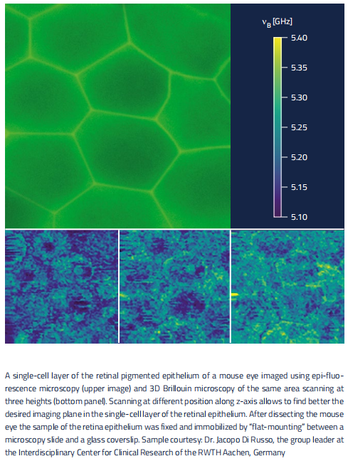
Subcellular-resolution 3D mechanical imaging of living tissues.
Suitable for mechanobiological research from single cells to whole small organisms (e.g., zebrafish embryos).
Applied in developmental biology, regenerative medicine, and tissue repair studies.
3. Plant Physiology
Real-time assessment of mechanical properties in plant tissues such as root tips and leaves.
Correlation with brightfield imaging to localize mechanical changes (e.g., cell wall thickening).
Supports studies on plant growth regulation, stress responses, and nutrient uptake mechanisms.
4. Soft Materials & Hydrogels
Quantitative analysis of stiffness and viscoelasticity in soft biomaterials such as hydrogels and Matrigel.
Ideal for evaluating scaffolds in tissue engineering, biomaterial development, and drug delivery platforms.
Allows in situ mechanical mapping of cell-laden hydrogels without additional sample preparation.
Product Advantages:
High-resolution and label-free imaging
Utilizing Brillouin microscopy technology, it enables non-invasive, label-free 3D mechanical property measurements of living cells, organoids, tissues, and small organisms.
Automation and ease of use
Employs intelligent workflow software such as FlowPlanner™ and PerfectOverlay™ to simplify operation, allowing users with little to no Brillouin microscopy experience to quickly obtain high-quality data.
Multimodal imaging integration
Seamlessly integrates with major inverted or upright microscopes, enabling simultaneous use with brightfield, phase contrast, DIC, fluorescence, and confocal imaging to perfectly combine mechanical information with optical imaging.
Strong compatibility with environmental control
Supports various environmental control devices, suitable for long-term live cell imaging experiments to ensure samples are measured under optimal conditions.
High sensitivity and stability
Uses a low-phototoxicity 780 nm near-infrared laser to minimize sample damage; the system is designed with mechanical and thermal stability for long-term experiments.
Powerful automation features
Includes automated calibration, autofocus, and large-area mapping (MultiMap™), reducing human error and improving data reliability and experimental efficiency.
Compatible with major microscope brands
Fits with mainstream microscopes such as Zeiss, Leica, Nikon, and Olympus, making it easy to integrate into existing experimental platforms.
Main Product Features:
1. Label-Free, Non-Invasive Imaging
Utilizes Brillouin scattering to measure mechanical properties without fluorescence labeling or damaging live samples.
2. 3D Mechanical Imaging Capability
Offers high-resolution 3D mechanical mapping of cells, organoids, tissues, and small organisms.
3. Highly Automated and User-Friendly
Equipped with intuitive software like FlowPlanner™ and PerfectOverlay™, enabling easy setup and operation—even for beginners.
4. Strong Multimodal Compatibility
Seamlessly integrates with brightfield, fluorescence, DIC, and confocal imaging for correlated optical and mechanical data.
5. Environmental Control Support
Compatible with temperature, CO₂, and humidity control systems for long-term live imaging applications.
6. High Sensitivity and Low Phototoxicity
Uses a 780 nm near-infrared laser to minimize photodamage, ideal for long-term observation of sensitive samples.
7. Advanced Data Processing and Visualization
Includes powerful features such as data fitting, edge detection, 3D rendering, and cross-sectional analysis, with batch processing support.
8. Modular Design with Broad Microscope Compatibility
Easily integrates with leading inverted and upright microscopes from Zeiss, Nikon, Leica, and Olympus, allowing easy system upgrades.
Application Areas:
1. 3D Cell Culture & Mechanobiology
Non-invasive, label-free measurement of the mechanical properties of spheroids and organoids.
Real-time monitoring of tumor organoids (e.g., MCF‑7 breast cancer) within 3D microenvironments.
Analysis of how cells respond to different mechanical conditions in terms of invasiveness, proliferation, and differentiation.
Investigation of time-dependent mechanical changes in organoid-based disease models.
2. Complex Tissue Mechanics
Subcellular-resolution 3D mechanical imaging of living tissues.
Suitable for mechanobiological research from single cells to whole small organisms (e.g., zebrafish embryos).
Applied in developmental biology, regenerative medicine, and tissue repair studies.
3. Plant Physiology
Real-time assessment of mechanical properties in plant tissues such as root tips and leaves.
Correlation with brightfield imaging to localize mechanical changes (e.g., cell wall thickening).
Supports studies on plant growth regulation, stress responses, and nutrient uptake mechanisms.
4. Soft Materials & Hydrogels
Quantitative analysis of stiffness and viscoelasticity in soft biomaterials such as hydrogels and Matrigel.
Ideal for evaluating scaffolds in tissue engineering, biomaterial development, and drug delivery platforms.
Allows in situ mechanical mapping of cell-laden hydrogels without additional sample preparation.
Detailed Specifications
Specification
Basic Configuration
Laser module
Spectrometer module
OptiBridge™ module
PC and software
System Specifications
Integrated closed-box system, with highest mechanical and thermal stability
Advanced fiber coupling to existing inverted or upright light microscopes
Near IR laser excitation with 780 nm for lowest phototoxicity and uncompromised fluorescence
Spectral resolution < 1 pm
Highest detector sensitivity for fastest signal capture
Transmission illumination with standard condensers for precise brightfield, DIC, and phase contrast
Fully automated system setup and calibration
Dimensions of laser module and spectrometer module (both stackable): 110 × 65 × 24 cm³
Dimension of OptiBridge™: 42×40×15 cm³
Optical Configurations
Fits on inverted motorized microscopes from:
Zeiss (Axio Observer line)
Olympus/Evident (IXplore line)
Nikon (Ti2 line)
Leica (DMi line)
Compatible with upright motorized fluorescence microscopes combined with Brillouin from:
Zeiss (Axio Imager)
Olympus/Evident (BX)
Leica (DM line)
Nikon (Ni line)
Brillouin simultaneously with other light microscopy methods
Fully simultaneous operation with optical phase contrast and DIC using standard condensers
Combine Brillouin with advanced commercial confocal microscopes and all advanced fluorescence optical techniques
Stages and Sample Holders
Compatibility with motorized stages is available for all major inverted optical microscope manufacturers such as Zeiss, Nikon, Olympus/Evident, and Leica
New Workflow-Based Discoverer™ Software
Revolutionary user experience with the new interface design
True multi-user platform, perfect for imaging facilities
New PerfectOverlay™ for combined optical and Brillouin or other information
MultiMap™ for multiple area mapping
FlowPlanner™ for designing a specific measurement workflow
Powerful Data Processing (DP) with full functionality for:
Data export
Fitting
Filtering
Edge detection
3D rendering
Cross-sectioning
Powerful batch processing of images and other analyses
Standard Operating Modes: Brillouin Operation
Brillouin mapping with Brillouin shift and linewidth
Advanced stiffness and viscosity mapping
PerfectOverlay™ for combining Brillouin and other optical images
Optical Imaging Modes
Brightfield imaging
Basic widefield fluorescence
Optional Modes and Accessories
FlowControl™ feature for remote experiment control
Wellplate mapping function
Epi-Fluorescence import function
Environmental control options
Additional XY or Z sample movement stages available


 Scan QR Code & Subscribe