
logo
×

PRODUCTS

Hydrogen-Induced Polarization MRI
  • You Are Here :HOME > PRODUCTS > Hydrogen-Induced Polarization MRI >
  • Hydrogen-Induced Polarization MRI

    description: HP metabolic magnetic resonance imaging technology enables real-time assessment of the kinetic behavior of cellular metabolism, thereby providing quantitative measurements of metabolic processes.
    Applications of MRI Polarization Equipment
    MRI (Magnetic Resonance Imaging) polarization equipment is primarily used to enhance the spin polarization of imaging targets (such as hydrogen nuclei or inert gases), thereby increasing the MRI signal strength. This enhancement improves image quality and diagnostic accuracy, expanding the boundaries of MRI technology. The following outlines its applications across various fields:
    I. Medical Diagnosis and Clinical Research
    1. Early Disease Diagnosis
    In the diagnosis of neurodegenerative diseases such as Alzheimer’s disease (AD), MRI polarization equipment can amplify signals from specific molecules in brain tissue. By using hyperpolarized water or specific metabolites (e.g., hyperpolarized pyruvate) as tracers, polarization equipment increases their polarization levels. After intravenous injection, MRI imaging can be used to monitor brain metabolic changes in real-time, allowing the detection of subtle abnormalities before clinical symptoms appear—enabling early diagnosis.
    2. Cardiovascular Disease Assessment
    For myocardial infarction patients, MRI polarization equipment combined with hyperpolarized gases (such as ¹²⁹Xe) enables clear visualization of myocardial ventilation and perfusion. After polarization, ¹²⁹Xe achieves a high polarization rate; when inhaled, it effectively labels pulmonary gas exchange, reflecting both cardiac function and pulmonary blood flow. This provides a solid basis for precise cardiovascular disease evaluation.
    3. Tumor Metabolism Analysis
    Tumor cells exhibit high metabolic activity and increased uptake of nutrients such as glucose. Hyperpolarized glucose analogs prepared using MRI polarization equipment can be injected into the body to observe tumor uptake and metabolic differences via MRI imaging. This helps distinguish between benign and malignant tumors, evaluate treatment efficacy, and predict prognosis—supporting personalized treatment planning with data.
    II. Drug Development and Clinical Trials
    1. Drug Efficacy Evaluation
    In drug development, MRI polarization equipment enables real-time monitoring of a drug’s distribution, metabolism, and mechanism of action in vivo. For example, hyperpolarized pyruvate can be used as a tracer—after polarization and injection into animal models or humans, MRI imaging tracks its conversion into lactate, helping assess how a drug affects cellular metabolism and identify its targets. This accelerates drug discovery.
    2. Drug Safety Evaluation
    MRI imaging using polarization equipment can detect potential drug-induced structural or functional damage to organs (e.g., liver, kidneys) in animal models or human subjects. Hyperpolarized tracers enhance tissue signal contrast, helping researchers detect subtle lesions or metabolic abnormalities early, thereby assessing drug safety and reducing clinical trial risks.
    III. Materials Science and Industrial Applications
    1. Material Structure and Performance Research
    In the study of polymer and nanomaterials, MRI polarization equipment can analyze internal structures and molecular dynamics. By polarizing specific nuclear spins (e.g., ¹H, ¹³C), it enhances MRI signal intensity, enabling the acquisition of data such as pore structures, molecular chain alignment, and diffusion coefficients—guiding performance optimization and the design of new materials.
    2. Industrial Non-Destructive Testing
    For materials like metals and ceramics, MRI polarization equipment used with suitable tracers can perform non-destructive internal defect detection (e.g., cracks, pores). By enhancing the signal through polarization, imaging resolution improves, enabling the detection of micron-level defects—ensuring product quality and safety in fields such as aerospace and mechanical manufacturing.
    IV. Fundamental Biomedical Research
    1. Cellular Metabolism Studies
    In cell biology, MRI polarization equipment can be used to observe dynamic changes in intracellular metabolites. By introducing hyperpolarized metabolites into cells and using MRI imaging to trace their metabolic pathways and rates, researchers can study core biological processes such as energy metabolism and signal transduction, and uncover molecular mechanisms behind disease development.
    2. Biomolecular Interaction Research
    For interactions such as protein-ligand and DNA-drug binding, MRI polarization equipment combined with hyperpolarized tracers enables real-time monitoring of molecular binding events and kinetics. Changes in hyperpolarized signal allow for the analysis of binding sites and affinities, providing critical data for drug design and target validation.
    HP metabolic MRl allows to assess in real-time the kinetic behavior of cellmetabolism-giving a quantitative measure of metabolism as it occurs.

    Real-time quantification of metabolic processes by means of HP MRI:
    Left:HP MRl allows the assessmentof metabolite uptake as wel as theidownstreamintracelular conversionincluding conversion to lactate.bicarbonate,and alanine.
    Middle:Conceptualyisualization ofmeasured signals for hyperpolarizedpyruvate and lactate and bicarbonateas downstream products of pyruvate.
    Right: Lactate/pyruvate ratio afterinjection of hyperpolarized pyruvate,coronal slice ofa subcutaneousMAT-B-lll breast cancer model (rat)
    surrounded by gel for B,homogeneity;3D dynamic bSSFP sequence.
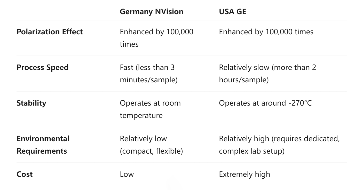
    HP metabolic magnetic resonance imaging (MRI) technology enables real-time assessment of the dynamic behavior of cellular metabolism. In clinical applications, according to research conducted by the Cambridge Cancer Center, this metabolic MRI technique has achieved a 90% accuracy rate in determining the efficacy of breast cancer treatment within seven days. The 13C pyruvate tracer used is a natural agent, posing no harm to the human body. In contrast, existing PET-CT technologies require approximately two months for evaluation and carry radiation risks. Competitive analysis indicates that GE Healthcare in the United States currently offers a similar product.


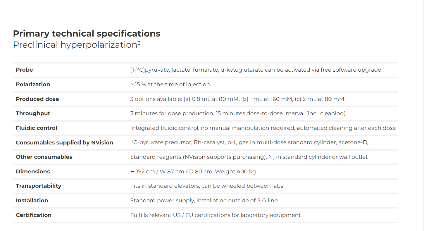
    Detailed Specifications