
logo
×

PRODUCTS

CellWave Sorter

  • You Are Here :HOME > PRODUCTS > CellWave Sorter >
  • CellWave Sorter

    description: CellWave Acoustic Microfluidics non-destructive cell sorting system -- a next-generation cell sorting platform based on acoustic microfluidics technology. Whether you are engaged in cell therapy, immunology research, oncology analysis, or rare cell captur
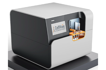
    CellWave Acoustic Microfluidic Label-Free Cell Sorting System
    CellWave is a next-generation cell sorting platform based on Acoustic Microfluidics technology. Whether you're working in cell therapy, immunology, oncology, or rare cell isolation, CellWave offers a label-free, non-invasive, high-purity cell sorting solution. It is a truly damage-free platform for cell sorting!
    Technical Advantages
    1.Non-invasive Sorting
    Cell sorting is achieved using acoustic waves, maintaining low shear force throughout the process and preserving the natural state of cells. It is particularly suitable for sensitive samples such as stem cells, extracellular vesicles (EVs), and primary cells.
    2.Ultra-high Purity and Recovery
    By precisely manipulating cells with acoustic standing wave fields, sorting purity exceeds 95%, and recovery rates exceed 80%—greatly surpassing traditional methods like FACS (Fluorescence-Activated Cell Sorting) or MACS (Magnetic-Activated Cell Sorting) in gentleness.
    3.High Throughput and Automation
    With a processing speed of up to 10⁶ cells/min, CellWave supports continuous sorting, minimizing manual errors. Integrated smart software allows one-click parameter setup, enabling easy and reproducible experiments.
    4.Versatile Compatibility
    Compatible with a variety of sample types, including blood, tissue dissociates, and cultured cells. Its modular design supports custom sorting strategies, such as sorting based on cell size, density, or mechanical properties.
    Technology Principle
    CellWave uses high-frequency ultrasound (MHz range) to generate a stable standing acoustic wave field inside a microfluidic chip. This allows non-contact manipulation of cells or particles through Acoustic Radiation Force:

    Standing Acoustic Wave Field
    Generated by a piezoelectric transducer, creating periodic high/low-pressure nodes inside the microchannel.
    Acoustic Radiation Force
    Cells are driven by pressure differentials in the acoustic field, migrating toward pressure nodes or antinodes, depending on their physical characteristics.
    Sorting Mechanism
    Cells of different sizes, densities, or mechanical properties respond differently to acoustic forces, enabling precise separation.
    Operation Workflow
    Sample Injection
    Cell suspensions flow through the microfluidic channel under laminar flow conditions.
    Acoustic Field Activation
    High-frequency ultrasound is activated, forming a standing wave field. Cells concentrate at pressure nodes.
    Sorting and Output
    Target cells: Deflected by acoustic forces and collected at the designated outlet.
    Non-target cells: Flow along the waste channel for removal.
    Gentle Recovery
    Sorted cells maintain high viability and are ready for direct downstream culture or analysis.
    Core Application Areas
    Cell Therapy and Regenerative Medicine
    Sorting of stem cells (e.g., CAR-T, iPSC preparation) to ensure the safety and consistency of clinical-grade cell products.
    Cancer Research and Liquid Biopsy
    Efficient enrichment of circulating tumor cells (CTCs) and extracellular vesicles, supporting early cancer screening and treatment monitoring.
    Immunology and Infectious Diseases
    Sorting of rare immune cells (e.g., antigen-specific T cells), accelerating vaccine development and immune response studies.
    Microbiology and Synthetic Biology
    High-throughput sorting of bacteria and yeast, optimizing strain selection and biomanufacturing processes.

    Advantages Compared to Other Sorting Technologies

    Parameters

    CellWave sound wave sorting

    Conventional Flow Sorting (FACS)

    Magnetic bead sorting (MACS)

    Cell damage

    no

    Laser/voltage damage cells

    Antibody labeling may activate the cells

    Labeling requirements

    Not required (can be marked if the sample has special requirements)

    Fluorescently labeled

    Magnetic bead labeling is required

    Flux

    High (continuous flow)

    High (Single cell detection)

    Low (Batch processing)

    Cost-effectiveness

    Low initial purchase cost and simple later maintenance

    The initial purchase cost is high, and the later maintenance cost of the laser is high

    The initial purchase cost is high, and the later use cost of the magnetic beads is high


    Detailed Specifications


    Classification

    Parameters (English)

    Parameters (Chinese)

    Specifications

    Optical system

    Laser

    Laser wavelength

    488 nm


    Fluorescence Filter

    Fluorescence filter

    530/43 nm


    Detectors

    Detector

    FITC (Fluorescence), FSC (forward scattering)

    Fluid systems

    Sample input


    Maximum Input Sample Volume

    Maximum sample volume

    600 µL


    Minimum Input Sample Volume

    Minimum sample volume

    150 µL


    Recommended Input Volume

    Recommended sample volume

    150-300 µL


    Recommended Concentration

    Recommended sample concentration

    0.5-1.5×10⁶ cells/mL


    Maximum Sheath Volume

    Maximum sheath volume

    1.2 mL


    Max Sheath Pressure

    Maximum sheath fluid pressure

    5 PSI


    Recommended Sheath Pressure

    Recommend sheath pressure

    2.25 PSI

    Output and Performance

    Output Containers

    Output containers

    2×1.5 mL EP tubes


    Throughput

    Flux

    10,000 cells/sec


    Max Sample Pressure

    Maximum sample pressure

    2 PSI


    Recommended Sample Pressure

    Recommended sample pressure

    1 PSI