

logo
×

PRODUCTS

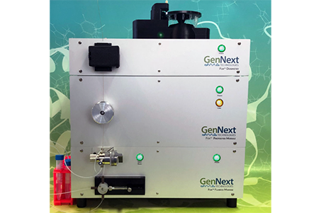
Fox Flash Oxidation Protein Footprinting System
  • You Are Here :HOME > PRODUCTS > Fox Flash Oxidation Protein Footprinting System >
  • Fox Flash Oxidation Protein Footprinting System

    description: GenNext has pioneered a superior, compact, cost-effective, and safe means of performing Hydroxyl Radical Protein Footprinting (HRPF) analysis by replacing expensive, complicated, and hazardous lasers with our proprietary Flash Oxidation (Fox™) Protein Foo
    Product Overview
    The Fox™ Photochemical Protein Footprinting System is a superior, compact, cost-effective, and safe solution for hydroxyl radical protein footprinting (HRPF). It replaces expensive, complex, and hazardous laser-based platforms with our proprietary Fox™ desktop rapid oxidation system.
    To address challenges in reproducibility, GenNext developed the Fox Radical Dosimeter—the world’s first system for measuring OH radicals. It automatically regulates the generation of effective OH radicals in the presence of unpredictable and varying background scavengers. The dosimeter enables real-time measurement of OH radical concentration, simplifying the assessment of biopharmaceutical solutions or experimental conditions prior to MS analysis. This significantly enhances HRPF usability while ensuring labeling credibility and fidelity.
    To eliminate today’s bottlenecks in bioinformatics and data processing, GenNext introduced FoxWare™, a unique software suite specifically designed to automate and accurately process data for HRPF higher order structure (HOS) analysis.
    Unique Advantages of the Fox System
    Direct Assessment of Solvent Accessibility
    Insights gained from studying various classes and systems of proteins using the Fox footprinting method can significantly influence decision-making at every stage of the drug discovery and development process—ultimately enabling the design and production of safer, more effective, and more stable biologics.
    Advanced Structural Validation
    The Fox system validates higher order structure (HOS) of complex biologics (such as monoclonal antibodies) by confirming correct folding and assembly. By comparing experimental Fox data with expected solvent accessibility derived from known protein structures, researchers can assess structural integrity and accuracy of biotherapeutics.
    High Spatial Resolution
    The Fox system offers high spatial resolution data about protein structure. It can precisely identify specific amino acids and regions that are solvent-exposed or buried. This level of detail allows researchers to map protein surfaces and accurately identify regions involved in protein–protein or protein–ligand interactions.
    Versatility Across Protein Classes
    The Fox system accommodates a wide range of protein classes, including soluble proteins, membrane proteins, and protein complexes. It is not limited by protein size, type, or structure, enabling the study of diverse systems. Its ability to handle very large and heterogeneous samples provides deep insights into their structure and functional dynamics.
    Complement to Other Techniques
    The Fox system complements other structural biology methods such as X-ray crystallography, NMR spectroscopy, hydrogen/deuterium exchange (HDX), and cryo-electron microscopy (cryo-EM). It provides information on solvent accessibility and protein dynamics in native solution-state conditions—data often inaccessible through other methods. When combined with these techniques, Fox-HRPF offers a more comprehensive understanding of protein structure and function.
    New Insights Enabled by the Fox System
    Protein–Protein Interactions
    The Fox® system identifies interaction sites and interfaces in protein–protein complexes. By measuring changes in solvent accessibility at interface residues upon binding, it can precisely locate the regions or domains responsible for interaction. This is particularly useful for studying interactions involving monoclonal antibodies and their antigens or other binding partners.
    Epitope Mapping
    Fox provides valuable information about solvent accessibility of residues involved in antigen–antibody interactions and reliably detects changes upon binding. This enables rapid identification and mapping of epitopes on monoclonal antibodies, offering insights into the binding interface and recognition regions. Such information is crucial for the design and optimization of therapeutic antibodies and diagnostic reagents.
    Characterization of Protein–Ligand Interactions
    The Fox system delivers key information on protein–ligand binding. By evaluating changes in solvent accessibility upon ligand binding, it helps identify residues at the binding site and reveals ligand-induced conformational changes. These insights support understanding of binding mechanisms and guide rational drug design.
    Protein Folding & Conformational Changes
    The Fox system detects conformational dynamics within proteins. By comparing Fox data across different conditions or time points, researchers can pinpoint structural rearrangements, as well as exposure or shielding of residues. This enables studies of conformational changes due to ligand binding or environmental factors.
    Allosteric Region Identification
    Fox identifies allosteric regions that are critical for understanding how proteins signal and undergo functional transitions. By revealing these regulatory sites, Fox-based HRPF facilitates elucidation of functional mechanisms and supports drug discovery targeting allosteric hotspots.
    Glycoprotein Analysis
    The Fox system reveals solvent accessibility in glycoproteins. Glycans can shield specific amino acids from oxidation, providing insights into glycan–protein interactions. Additionally, the method aids in understanding conformational shifts under various conditions and the structural basis of glycoprotein function.

    Figure 1: HRPF Principle Illustration — Fast Photochemical Oxidation of Proteins (FPOP)
    In FPOP, proteins are mixed with hydrogen peroxide and exposed to pulsed UV light, which photolyzes H₂O₂ into •OH radicals that oxidize solvent-exposed amino acids. After labeling, samples are quenched in a solution containing catalase and methionine, digested, and analyzed using high-resolution LC-MS/MS.
    Application Areas
    Monoclonal Antibodies
    Epitope Mapping
    Paratope Mapping
    Aggregation Studies
    Formulation Research
    Biosimilar Development
    Originator Comparison
    Drug Target Discovery
    Drug Binding Site Characterization
    Allosteric Effects
    Biomolecular Interactions
    Protein–Ligand
    Protein–Protein
    Detailed Specifications