

Single-cell spatial protein tissue MS imaging system -MIBIscope system
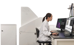
Single-cell spatial protein tissue MS imaging system -MIBIscope system |
|
| description: | MIBIscope, which combines spatial imaging methods with mass spectrometry, may help achieve another dimension in precise imaging of the tumor microenvironment. It uses metal isotope elements as reporter tags to label antibodies and simultaneously detect up |
|---|---|
1. A disruptive multi-tissue imaging platform—visualize more than 40 markers in a single image
Multiple ion beam tissue mass spectrometry imaging instrument is applied to high-precision single-cell spatial proteomics. Based on multiple ion beam imaging (MIBI) technology, the MIBIscope system can visualize 40+ protein markers in a single scan, and provide tissue sample microenvironment. Related Information. The system can display more than 40 markers simultaneously with single-step staining and single-step imaging. MIBIscope images from 3mm x 3mm scans of granulomatous lung sections from patients infected with Mycoplasma. Left panel shows immune infiltration in infected tissue (CD45, yellow; CD31, red; SMA, blue; CD68, magenta). magnified single
2. High resolution - achieve confocal resolution
MIBIscope can be adjusted like a light microscope, with resolution settings from 280 nm to 1 µm. At higher magnifications, MIBIscope provides resolution comparable to confocal microscopy.
3. High sensitivity - single molecule sensitivity
MIBIscope uses highly sensitive secondary ion mass spectrometry, allowing single-molecule detection, as demonstrated in Science Advances and other recent publications.
feature of product:
(1) MIBI is compatible with all common sample types, including fresh frozen and FFPE tissue. If you have an antibody that works by traditional IHC, we'll likely be able to validate it for MIBI.
(2) All imaging is done using the proprietary MIBIscope™ system, we can image ten slides or hundreds of samples, all overseen by a board certified pathologist, ensuring consistency throughout data acquisition sex.
(3) Our data science team analyzes the images to gain insight into the tumor microenvironment, with standard analyses including comprehensive enumeration of immune cell populations, single-cell-based quantification of checkpoint marker expression, and spatial analysis. For example, we can quantify the proximity of effector cells (e.g. PD-1+, CD8+ T cells) to target cells (e.g. proliferating PD-L1+ tumor cells) and generate a number of similar statistics to distinguish responders from non-responders By.
concrete application:
With MIBIscope™, scientists can analyze multiple markers simultaneously using a single slide. Expression can be measured at the single-cell level with subcellular resolution while preserving spatial information.
In a study recently published in Cell, multiplexed ion beam imaging revealed a structured tumor immune microenvironment in triple-negative breast cancer, Keren et al. Comprehensive profiling of the tumor microenvironment in 41 triple-negative breast cancer patients using MIBI technology. The authors simultaneously imaged 36 proteins at subcellular resolution in diagnostic surgical pathology samples.
Using a multi-step analysis including cell segmentation, single-cell analysis, and spatial analysis, the authors reveal how tumor expression and immune components correlate in the tissue context, and how this correlates with overall survival in TNBC. The authors show how to use the MIBI™ multiplexed imaging approach to stratify patient populations and identify unique patient populations that respond to treatment.

 Scan QR Code & Subscribe